Post Time:Nov 22,2011Classify:Company NewsView:514
Carl Zeiss Microscopy, a company of the Carl Zeiss Group and leading provider of light, laser-scanning and electron and ion beam microscopes, today announced the celebration of its tenth anniversary of the CrossBeam® FIB-SEM technology.
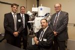
In honor of the tenth anniversary, a ceremony was held at the Center for Composite Materials at the University of Delaware, who have acquired an AURIGA 60 CrossBeam workstation. The system is the first major acquisition intended for the University´s new Interdisciplinary Science and Engineering Laboratory (ISE Lab), now under construction. When completed, the ISE Lab will house their new AURIGA 60 CrossBeam FIB-SEM instrument and provide nearly 200,000 square feet of space for hands on research and education. According to Dr. David Martin, Chair of Materials Science and Engineering, “This multipurpose analytical instrument is an excellent demonstration of the interdisciplinary research opportunities that will be provided by the ISE Lab. The microscope will be open to use by all members of the University of Delaware community, as well as our partners in industry, government laboratories and neighboring academic institutions.”
“The versatile, flexible AURIGA CrossBeam system has become the most successful FIB-SEM instrument ever produced by Carl Zeiss,” said Michael Rauscher, Director of the Product Segment CrossBeam. “We
Considered by users as the industry´s most versatile FIB-SEM workstation, the AURIGA instrument includes both a GEMINI® scanning electron microscope (SEM) column and a focused ion beam (FIB) column. The SEM column enables the AURIGA to create superb, high resolution, nano scale images, while the FIB column enables the tool to remove material from the sample by ion-milling. In fact, by automatically combining several images, a complete 3D-model of the sample can be created. Moreover the AURIGA can also be used to deposit material to the sample by means of various pre-cursor gases. These capabilities make the AURIGA ideally suited for today’s materials research with its great complexity of challenging tasks including high resolution imaging, chemical composition analysis, crystallographic orientation and electrical attributes. The tool is also superbly suited for life science research applications, e.g. in the growing field of brain-mapping. 
Source: Carl Zeiss MicroImaging GmbHAuthor: shangyi
PrevOnline Selection of 2011 Top 10 Brand of Glass Industry has Started
International company to locate U.S. operation in LavoniaNext